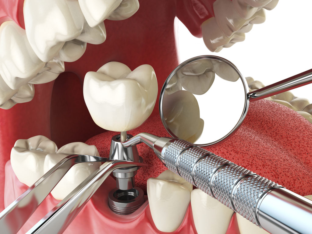
Guided implant surgery is one more way to make the dental implant process even more accurate.
Guided implant surgery includes the same steps and concepts as traditional implant surgery. However, by using CBCT and CAD/CAM technology, we can provide a restoratively driven approach that provides the best functioning implant while taking the guesswork out of the procedure — creating a more predictable and comfortable procedure for our patients.
The first step is to evaluate 3-D X-ray images taken through our CBCT (cone beam computed tomography) scanner. Rather than rough measurements on 2D X-rays and visual inspection of the area, an accurate 3-D image of the patient’s skeleton can be manipulated in the computer. Accurate measurements can be made, and the bone can be more precisely evaluated for quality and quantity. As a result, Dr. Fienman can determine any need for bone grafting at that point as well.
The implant can be placed in the site virtually. While evaluating for size, angulation, ideal positioning, and occlusion of the new crown, Dr. Fienman can better identify and avoid any vital structures like blood vessels and nerves that could complicate things on surgery day.

Utilizing this information and a digital impression of the patient’s mouth, a surgical guide/surgical stent can be designed and fabricated for use on the actual surgery day. A surgical stent provides Dr. Fienman with a tool to translate the exact location and angulation of the implant from the virtual surgery to real life. Utilizing the surgical stent makes the surgery day faster and easier for the patient.
After the implant has healed and is ready to be fit for a crown, rather than traditional goopy impressions, we can utilize special scan bodies and the digital impression to generate a 3-D printed model to be used by the lab.
All this technology means more predictability, accuracy, and comfort for our patients.
Call us for more information about guided implant surgery or to make an appointment with Dr. Fienman.


